Home
Portfolio
About
Blog
Contact Us
Search
Portfolio
All
Cardiovascular
Cellular
Dermatology
Embryology
Gastrointestinal
Medical Device
Musculoskeletal
Neurology
Obstetrics and Gynecology
Oncology
Orthopedic
Pharmacology
Surgical
Veterinary
Cardiovascular
,
Neurology
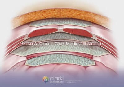
Carotid Artery Blockage
Gastrointestinal
,
Musculoskeletal
,
Surgical
Abdominal Hernia Graft Placement
Cardiovascular
,
Cellular
,
Pharmacology
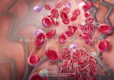
Dual Antiplatelet Therapy
Dermatology
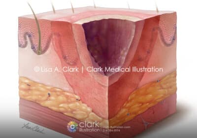
Burn Zones
Obstetrics and Gynecology
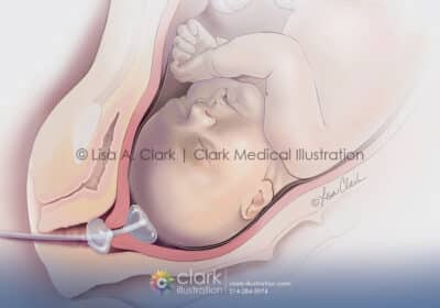
Cervical Ripening Balloon
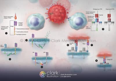
Cellular
,
Oncology
CAR-T and TCR Therapies for Cancer Treatment
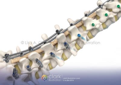
Medical Device
,
Musculoskeletal
,
Neurology
,
Orthopedic
Medtronic ModuleX Spinal System
Cellular
,
Neurology
,
Pharmacology
Alzheimer’s Markers in the Blood
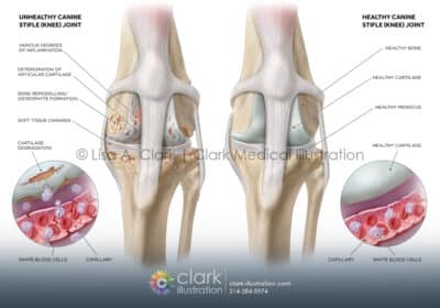
Orthopedic
,
Veterinary
Canine Knee (Stifle) Joint
Cellular
,
Pharmacology
Yeast and Neurodegenerative Disease Research
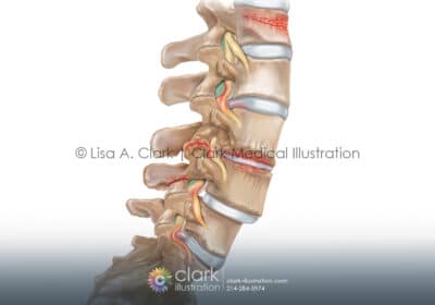
Musculoskeletal
,
Neurology
,
Orthopedic
,
Surgical
Spinal Conditions
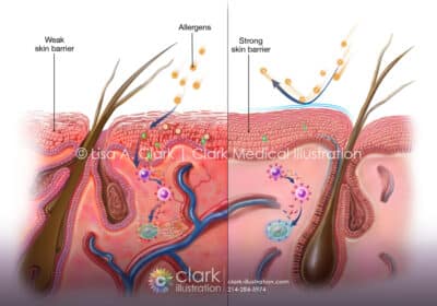
Cellular
,
Dermatology
,
Veterinary
Canine Skin Barrier and Allergies